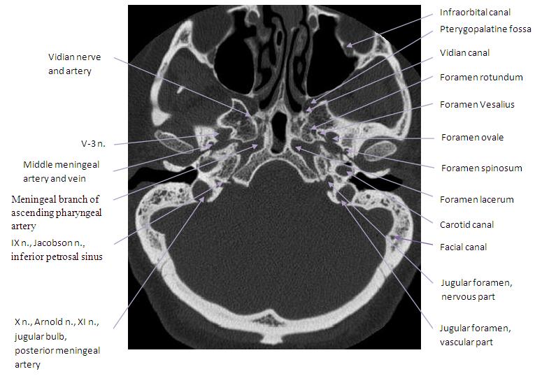
skull and brain anatomy
EPOS™ - C-0157 we have 9 Pictures about EPOS™ - C-0157 like Poster : Sagittal Section Of Brain in Situ, MRI Brain Anatomy | Radiology Anatomy Images | Mri brain, Brain anatomy and also MRI Brain Anatomy | Radiology Anatomy Images | Mri brain, Brain anatomy. Here you go:
EPOS™ - C-0157
 epos.myesr.org
anatomy skull ct base epos mri
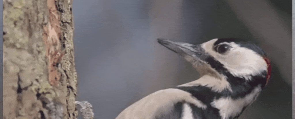
How Do Woodpeckers Survive Repeated High-Impact Shocks Without Brain
 www.sciencealert.com
brain pecker
Broken Human Skull Close Up. Toned Image. Royalty Free Stock
 www.pinterest.com
skull reference anatomy broken drawing
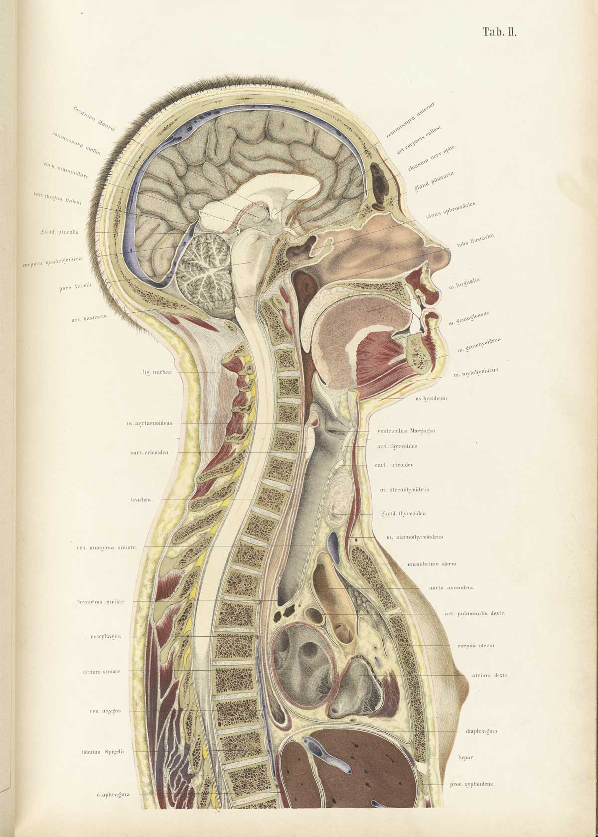
Historical Anatomies On The Web: Wilhelm Braune Home
 www.nlm.nih.gov
section cross head braune wilhelm human anatomy street body exhibition female atlas ii table anatomischer anatomies historical brain medical anatomical
Cerebrum Sheep Dissection - Human Anatomy - GUWS Medical
 www.guwsmedical.info
anatomy sheep human dissection kangaroo brain cerebrum half midsagittal section right figure guws medical
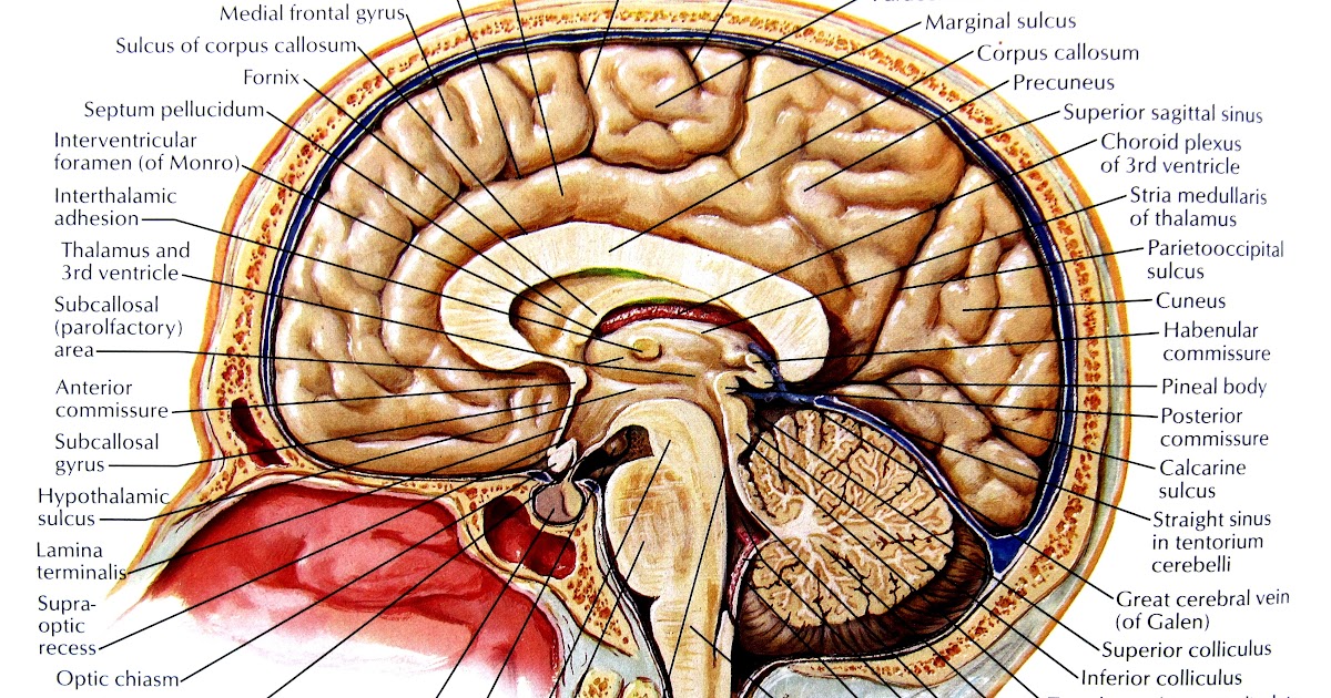
Poster : Sagittal Section Of Brain In Situ
 www.smallpocketlibrary.com
sagittal section situ
MRI Brain Anatomy | Radiology Anatomy Images | Mri Brain, Brain Anatomy
 www.pinterest.com
mri brain anatomy radiology labeled coronal brian labels imaging medical roi region mr normalized neuroanatomy sagittal rois volumetric illustration neuroscience
B Is For Basilar Suture; The Marker Of Fusion Of The Spheno-occipital
 www.pinterest.com
skull inferior basilar suture fusion occipital anatomy spheno synchondrosis human foramen physiology marker hn cranial nerves answers brain palatine flashcards
Passavant’s Ridge: Anatomy, Muscles And Clinical Aspects | Kenhub
stylopharyngeus muscle pharynx muscles anatomy kenhub constrictor ridge musculus pharyngeal superior inferior origin insertion innervation pars raphe passavant walls muskler
Passavant’s ridge: anatomy, muscles and clinical aspects. Anatomy skull ct base epos mri. Mri brain anatomy