pelvic female anatomy
USMLE CT SCAN ABD AND PELVIS - YouTube we have 9 Pictures about USMLE CT SCAN ABD AND PELVIS - YouTube like Pelvis - Learn Muscles, Interpreting X-Rays of the Pelvis, Hip Joint and Femur - YouTube and also Normal female pelvis, MRI - Stock Image - C026/9012 - Science Photo Library. Here it is:
USMLE CT SCAN ABD AND PELVIS - YouTube
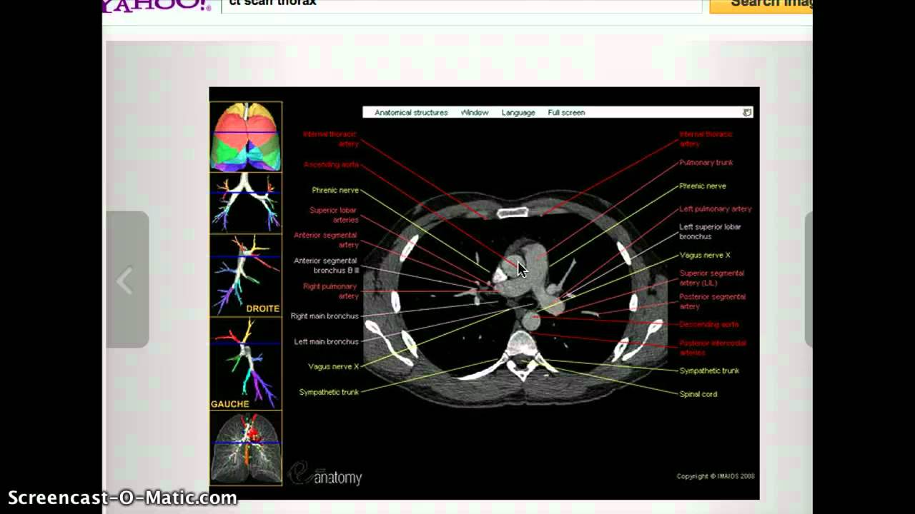 www.youtube.com
ct scan pelvis abd usmle
Pelvis - Learn Muscles
 www.learnmuscles.com
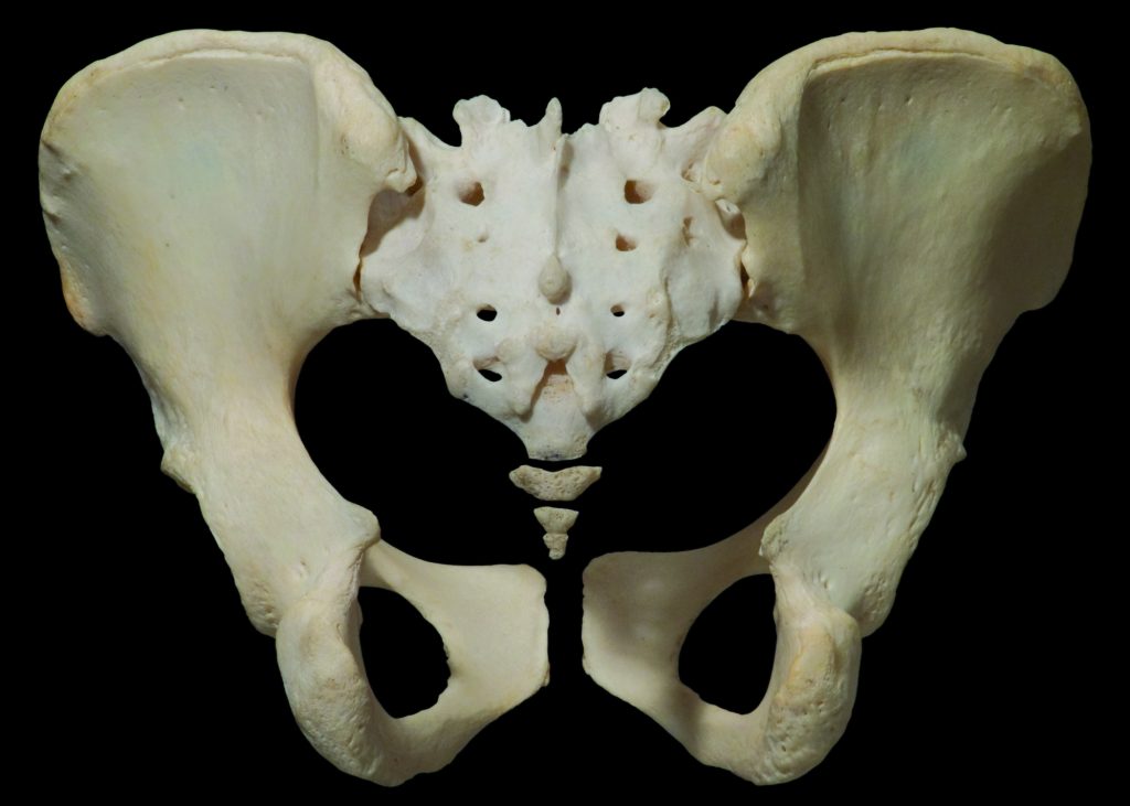
pelvis posterior bony anatomy joints female
Developmental Hip Dysplasia In Babies And Young Children
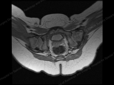 www.hss.edu
hip pediatric mri dysplasia developmental relocated children overview anatomically aligned hss edu
Small Intestine: Anatomy, Location And Function | Kenhub
intestine anatomy location function kenhub background
Pelvic Exercises - Pelvic Floor Safe Exercises For Women | Hysterectomy
 www.pinterest.com
exercises hysterectomy pelvic recovery floor surgery partial safe
Pelvis Computed Tomograph (axial CT)
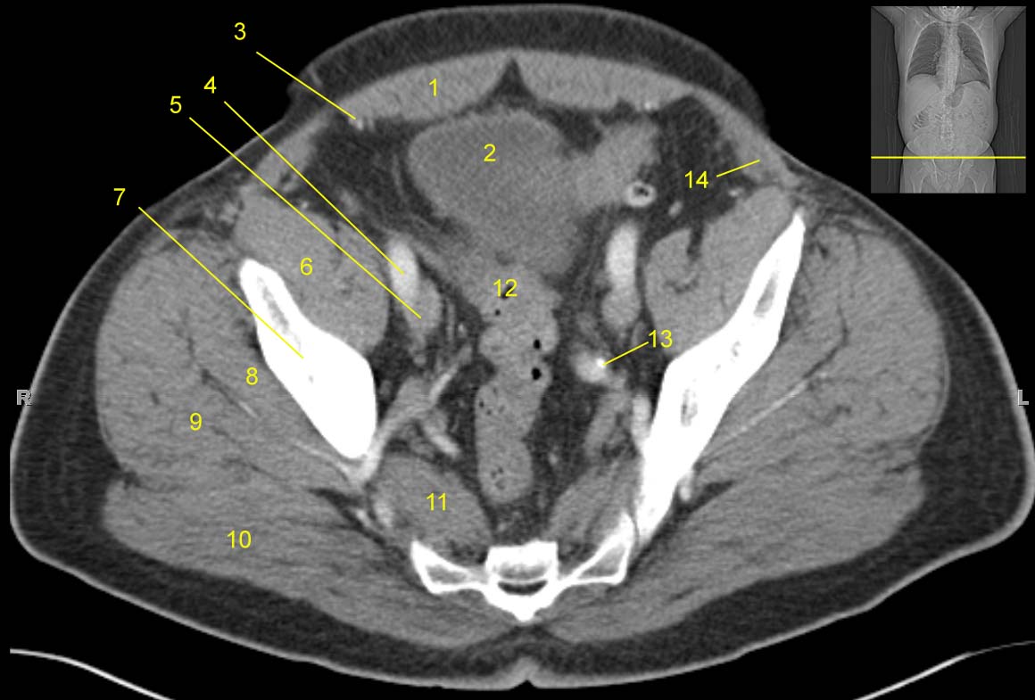 jhurads4anatomy.com
ct pelvis anatomy scan pelvic muscle axial iliacus bone labeled computed cat tomograph legend study ss1 docs storage google
Normal Female Pelvis, MRI - Stock Image - C026/9012 - Science Photo Library
 www.sciencephoto.com
mri pelvis female normal
Interpreting X-Rays Of The Pelvis, Hip Joint And Femur - YouTube
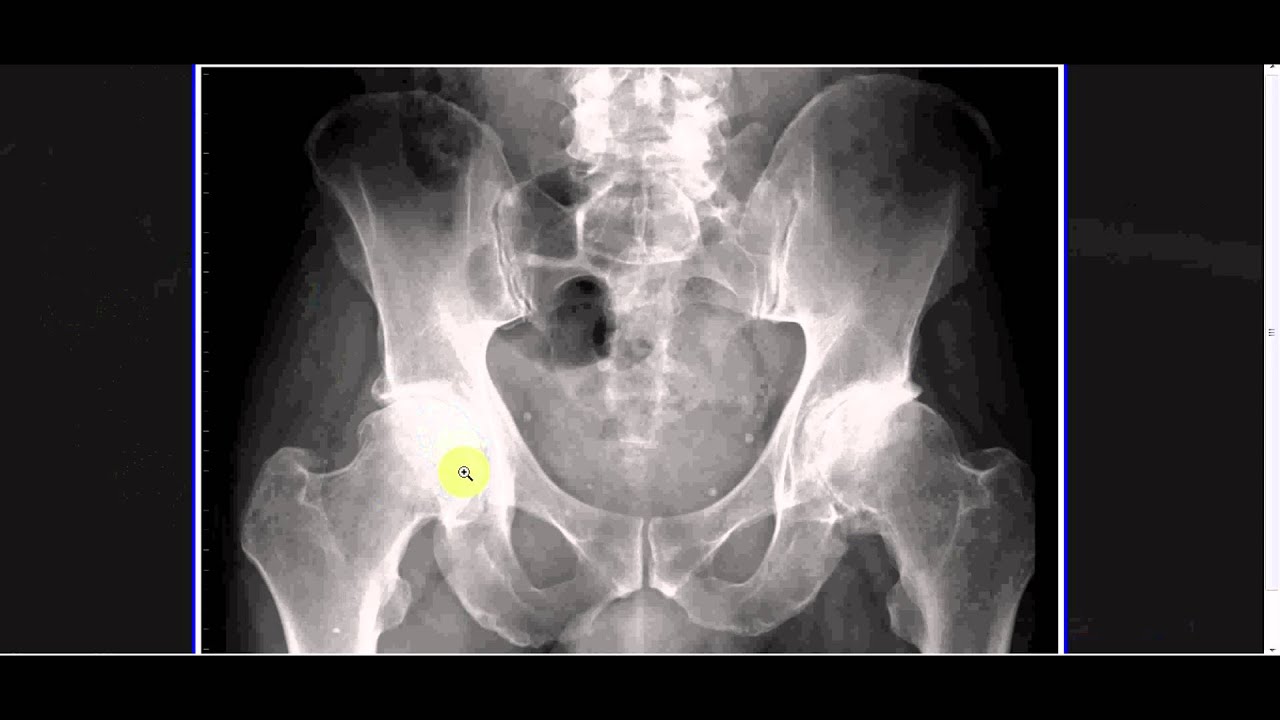 www.youtube.com
hip xray female joint pain pelvis femur interpreting rays anatomy pelvic arthritis bone ball
Diaphragm: Location, Anatomy, Innervation And Function | Kenhub
diaphragm intercostal spaces kenhub anatomy innervation muscles anterior vertebral location ventral
Interpreting x-rays of the pelvis, hip joint and femur. Pelvis posterior bony anatomy joints female. Hip xray female joint pain pelvis femur interpreting rays anatomy pelvic arthritis bone ball