neck artery anatomy
Transoral Lateral Oropharyngectomy for Squamous Cell Carcinoma of the we have 9 Pictures about Transoral Lateral Oropharyngectomy for Squamous Cell Carcinoma of the like Cross Section of Neck at the 7th Cervical Vertebra | ClipArt ETC, Perspective View of Left Lateral Neck | Neuroanatomy | The and also Left Lateral View of Frontal Lobe, Middle and Posterior Fossa Floors. Here it is:
Transoral Lateral Oropharyngectomy For Squamous Cell Carcinoma Of The
 jamanetwork.com
journals lateral neck jamanetwork
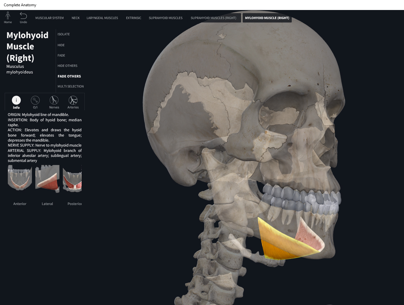
Muscles: Mylohyoid. – Anatomy & Physiology
 integrativewellnessandmovement.com
mylohyoid
Cross Section Of Neck At The 7th Cervical Vertebra | ClipArt ETC
neck section cross cervical vertebra 7th etc clipart tiff usf edu
Ultrasound Of The Pancreas: What Normal Looks Like
 www.auntminnie.com
ultrasound pancreas normal transverse sections image02 looks
Ulnar Nerve Subluxation: Clinical Anatomy | Kenhub
nerve median nervus medianus kenhub ulnar brachial muscle musculocutaneous coracobrachialis anatomy vein subluxation course ulnaris innervation clinical origin ventral plexus
Cervical Spine Ligaments | Radiology Reference Article | Radiopaedia.org
 radiopaedia.org
spine ligaments anatomy cervical radiology radiopaedia spinal vertebral ligament longitudinal flavum ligamentum apical supraspinous gaillard ortopedia thoracic medizin human fisiologia
Perspective View Of Left Lateral Neck | Neuroanatomy | The
 www.neurosurgicalatlas.com
neck left lateral neuroanatomy
Left Lateral View Of Frontal Lobe, Middle And Posterior Fossa Floors
 www.neurosurgicalatlas.com
fossa lateral posterior left lobe frontal floors middle neurosurgicalatlas
Foot, Plantar Surface (deeper) – Human Body Help
 www.humanbodyhelp.com
plantar foot surface deeper key
Ultrasound of the pancreas: what normal looks like. Left lateral view of frontal lobe, middle and posterior fossa floors. Journals lateral neck jamanetwork