diagram of gland
Where is the kidney? I feel pain in the lower side of abdomen we have 9 Pics about Where is the kidney? I feel pain in the lower side of abdomen like Diagram showing the relationship of the Parotid Gland & th… | Flickr, Wasp Anatomy and Diet | HowStuffWorks and also Diagram showing the relationship of the Parotid Gland & th… | Flickr. Here it is:
Where Is The Kidney? I Feel Pain In The Lower Side Of Abdomen
kidneys abdomen healthyandnaturalworld healthtopquestions
Diagram Showing The Relationship Of The Parotid Gland & Th… | Flickr
 www.flickr.com
parotid gland facial nerve anatomy salivary nerves neck swollen face removal branches diagram masseter dissection parotidectomy trigeminal cheeks tumor through
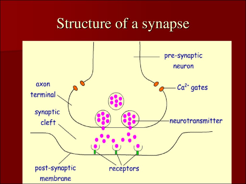
Nerve Centers. Synaptic And Junctional Transmission. Central Inhibition
 ppt-online.org
synapse structure synaptic transmission inhibition nerve ppt
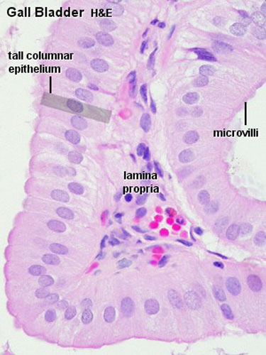
Gastrointestinal Tract - Gallbladder Histology - Embryology
 embryology.med.unsw.edu.au
histology gallbladder bladder cells gall tissue epithelium human embryology tissues duct bile normal liver tract pancreas atlas slides gastrointestinal cell
Coronal CT Image Showing The Calcified Mass Of 42 × 17 Mm In The Left
 www.researchgate.net
calcified submandibular gland coronal
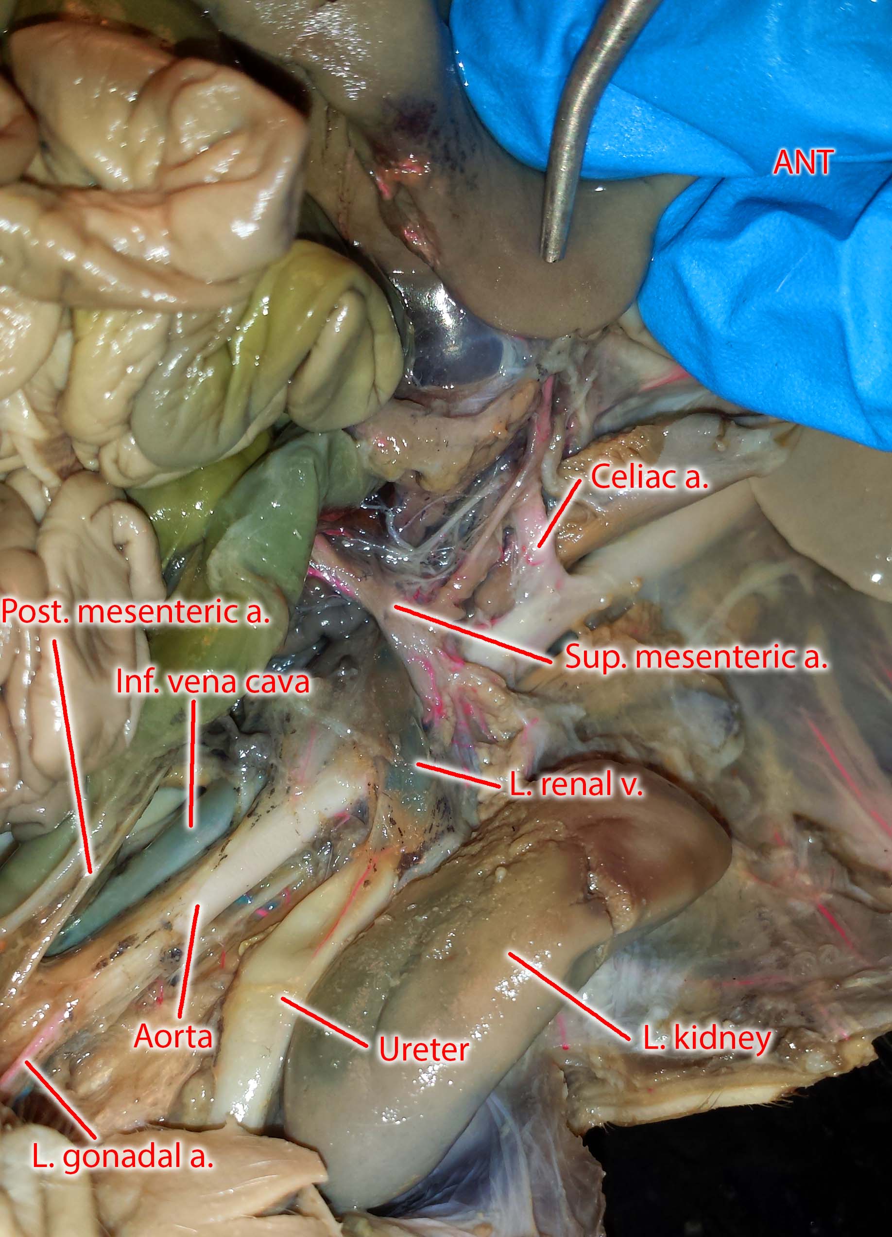
Photographs Of The Vessels Of The Fetal Pig
 blog.valdosta.edu
pig fetal artery mesenteric vein superior blood vessels renal vena cava vessel practical celiac inferior posterior stomach iliac umbilical common
Nervous System - Presentation Health And Disease
.PNG) www.sliderbase.com
www.sliderbase.com
nerve cells types main nervous biology control cell system structure basic humans unit source study hard
Integumentary System - Histology - Embryology
 embryology.med.unsw.edu.au
histology skin integumentary system dermis layers epidermis anatomy slides structures blood vessels hair physiology label cell diagram stain appendages draw
Wasp Anatomy And Diet | HowStuffWorks
 animals.howstuffworks.com
wasp anatomy wasps diet insects bees bee many howstuffworks honeybee sting stings
Photographs of the vessels of the fetal pig. Coronal ct image showing the calcified mass of 42 × 17 mm in the left. Nerve cells types main nervous biology control cell system structure basic humans unit source study hard