atlas anatomy bone
Osseous anatomy of the Sphenoid Bone | Neuroanatomy | The Neurosurgical we have 9 Images about Osseous anatomy of the Sphenoid Bone | Neuroanatomy | The Neurosurgical like Coxal (Pelvic) bone, lateral view with labels - Appendicul… | Flickr, Human Anatomy Video: The Typical Vertebra - YouTube and also Human Anatomy Video: The Typical Vertebra - YouTube. Here you go:
Osseous Anatomy Of The Sphenoid Bone | Neuroanatomy | The Neurosurgical
 www.neurosurgicalatlas.com
neuroanatomy
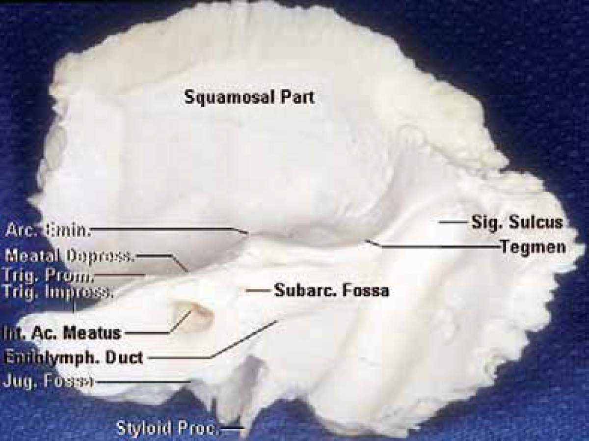
Oblique Posterior View Of Right Temporal Bone | Neuroanatomy | The
 www.neurosurgicalatlas.com
oblique neurosurgicalatlas correlation
Human Anatomy Video: The Typical Vertebra - YouTube
 www.youtube.com
vertebra typical anatomy human
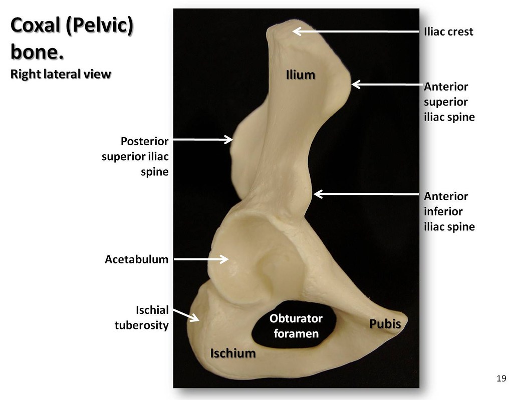
Coxal (Pelvic) Bone, Lateral View With Labels - Appendicul… | Flickr
 www.flickr.com
bone pelvic lateral coxal skeleton appendicular atlas anatomy labels visual bones flickr views pro flickriver
The Radiology Assistant : Temporal Bone - Anatomy 2.0 | Ear Anatomy
 www.pinterest.com
temporal nerve radiology attic eustachian imaging dysfunction istant
Neurovasculature Of The Cranial Fossa | Neuroanatomy | The
 www.neurosurgicalatlas.com
cranial fossa neurovasculature neuroanatomy correlation surgical
Superior View Of The Temporal Bone And Infratemporal Fossa And Orbit
 www.neurosurgicalatlas.com
fossa temporal infratemporal neuroanatomy
Anatomy Lab Photographs Vertebrae
 faculty.sdmiramar.edu
axis anatomy vertebrae c2 skeletal lab memrise faculty
Shoulder: MRI, Radiographical, And Illustrated Anatomical Atlas
 www.imaios.com
shoulder anatomy mri joint anatomical muscles ligaments labeled humeral gleno deep scapulohumeral imaios dissection biceps medical layer showing glenoid illustrations
Bone pelvic lateral coxal skeleton appendicular atlas anatomy labels visual bones flickr views pro flickriver. Shoulder anatomy mri joint anatomical muscles ligaments labeled humeral gleno deep scapulohumeral imaios dissection biceps medical layer showing glenoid illustrations. Axis anatomy vertebrae c2 skeletal lab memrise faculty