anatomy of brainstem
Maxilla: Anatomy, function, clinical aspects | Kenhub we have 9 Pics about Maxilla: Anatomy, function, clinical aspects | Kenhub like Neuro Lab Block I- Brain Stem Gross Anatomy Flashcards | Easy Notecards, Ventral View of Brainstem and Basal Forebrain | Neuroanatomy | The and also Posterior View of the Suboccipital Surface of the Cerebellum. Here it is:
Maxilla: Anatomy, Function, Clinical Aspects | Kenhub
maxilla kenhub anatomy function
Kidney: Blood Supply, Innervation And Lymphatics | Kenhub
kidney supply renal cortex innervation blood kenhub anatomy
Ventral View Of Brainstem And Basal Forebrain | Neuroanatomy | The
 www.neurosurgicalatlas.com
ventral forebrain brainstem neuroanatomy
Neuro Lab Block I- Brain Stem Gross Anatomy Flashcards | Easy Notecards
 www.easynotecards.com
anatomy peduncle brain stem cerebellar gross inferior superior middle neuro lab block easynotecards
Midline Deep Brain Anatomy And Pineal Region | Neuroanatomy | The
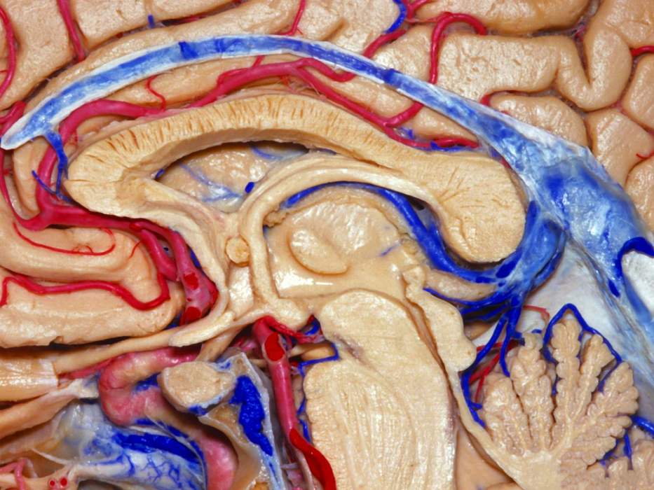 www.neurosurgicalatlas.com
brain anatomy pineal midline deep rhoton region aans human ventricle 3d atlas third structure gland correlation surgical livescience neurosurgicalatlas
Optic Neuropathy Due To Optic Compression: Clinical Case | Kenhub
willis circle optic nerve cadaver compression anatomy kenhub brain neuropathy base shows clinical due case
Neuroanatomy Labelled Specimen For 2nd Yr MBBS Spotting
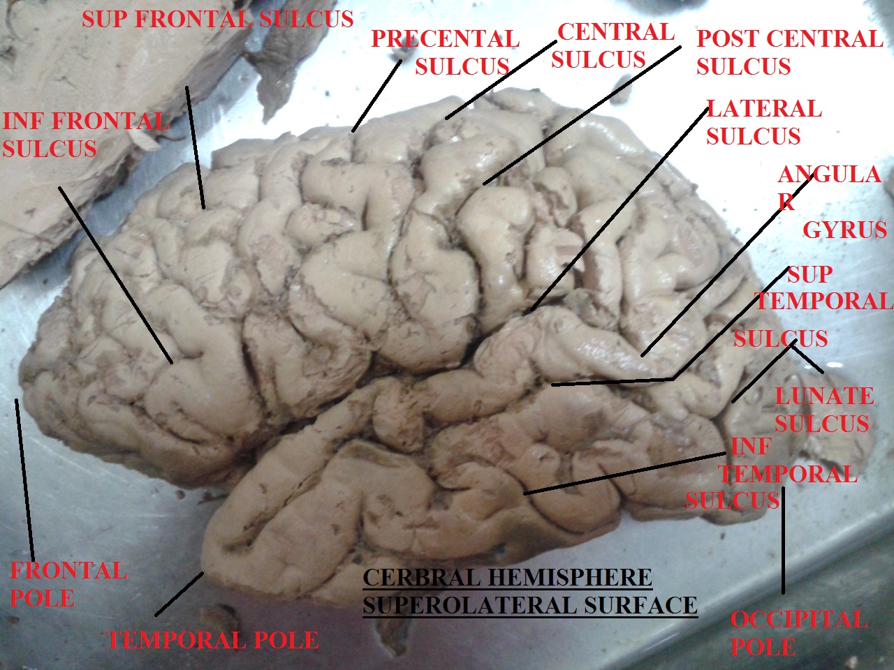 www.kemunited.com
specimen neuroanatomy superolateral hemisphere labelled neurology kemunited
Tentorial Incisura | Neuroanatomy | The Neurosurgical Atlas, By Aaron
 www.neurosurgicalatlas.com
tentorial incisura neuroanatomy rhoton surgical correlation
Posterior View Of The Suboccipital Surface Of The Cerebellum
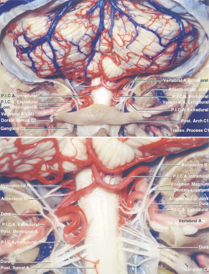 www.neurosurgicalatlas.com
cerebellum suboccipital posterior neuroanatomy rhoton surgical neurosurgicalatlas
Willis circle optic nerve cadaver compression anatomy kenhub brain neuropathy base shows clinical due case. Midline deep brain anatomy and pineal region. Neuroanatomy labelled specimen for 2nd yr mbbs spotting